Medial Collateral Ligaments Of The Knee / A Focus On Mcl Injuries Inline Physio : Athletes who participate in direct medial collateral ligament tears often occur as a result of a direct blow to the outside of the knee.
Get link
Facebook
X
Pinterest
Email
Other Apps
Medial Collateral Ligaments Of The Knee / A Focus On Mcl Injuries Inline Physio : Athletes who participate in direct medial collateral ligament tears often occur as a result of a direct blow to the outside of the knee.. The medial collateral ligament (mcl) and the lateral collateral ligament (lcl). These paired ligaments are called the anterior cruciate ligament (acl) and the. The medial collateral ligament (also known as tibial collateral ligament) is a strong, flat band on the medial element of the knee joint. The medial collateral ligament (mcl) is found on the side of the knee closest to the other knee. It has superficial and deep portions.
The medial collateral ligament (mcl) is the most important stabilizer of the medial side of the knee together with the capsuloligamentous complex. J bone joint surg am. The medial collateral ligament (also known as tibial collateral ligament) is a strong, flat band on the medial element of the knee joint. Related online courses on physioplus. The medial supporting structures are the most commonly injured ligaments in the knee.
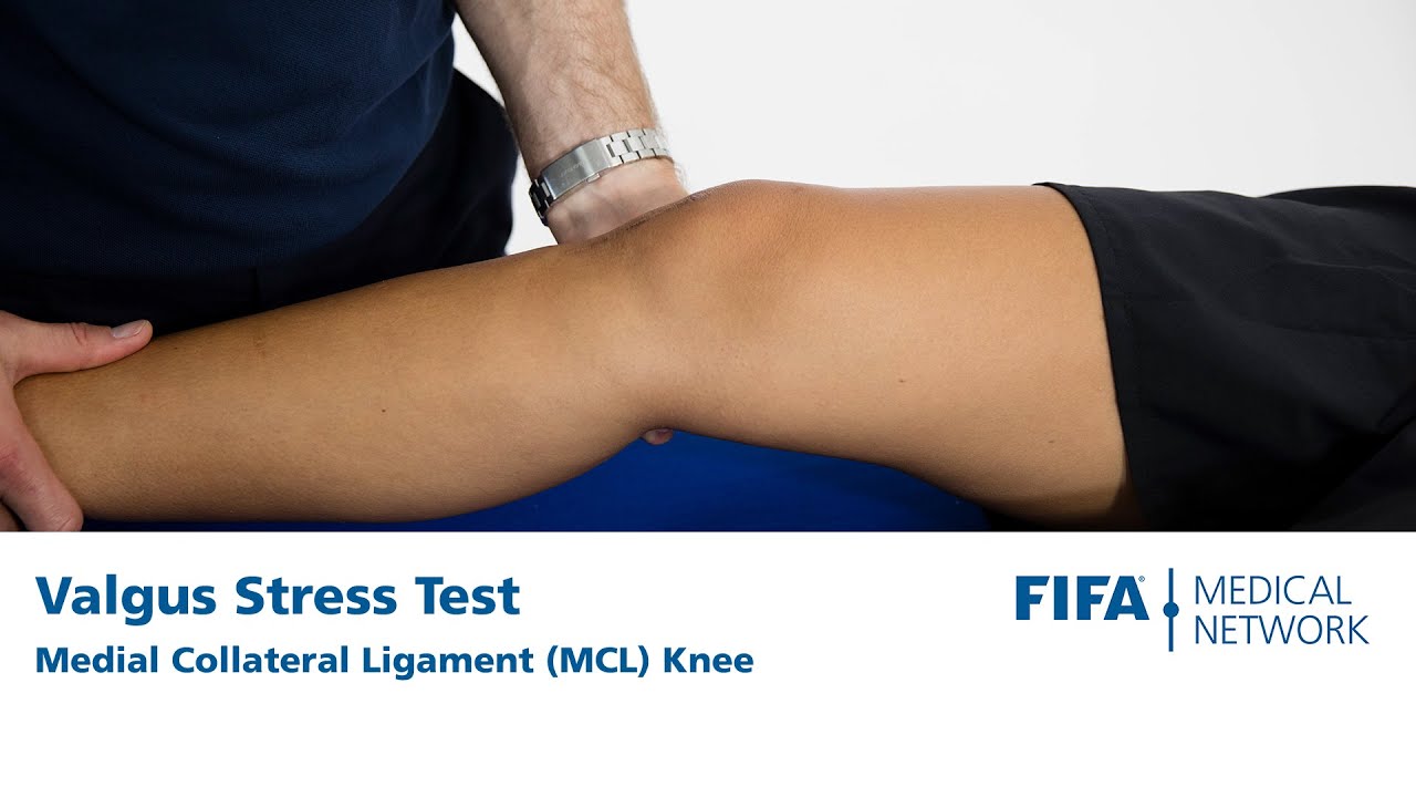
Valgus Stress Test Medial Collateral Ligament Mcl Knee Youtube from i.ytimg.com The knee has 2 collateral (parallel) ligaments and 2 cruciate (crossing) ligaments. This stretches the ligaments on the inside of the knee too far or can tear them. An example of this is when. It is also a secondary restraint to for patients with medial collateral ligament injuries, it is important to document the precise location of the tenderness. It is a thick fibrous band and is designed to resist valgus the onset is usually traumatic and can happen via a direct blow to the outside of the thigh. The medial collateral ligament (mcl) runs along the inside of your knee. Courtesy of randale sechrest, md, ceo, medical multimedia group. There are two collateral ligaments of the knee:
The medial collateral ligament (mcl) is the most frequently injured structure of the knee.
The knee has very little inherent bony stability: The medial collateral ligament (mcl) and lateral collateral ligament (lcl) serve as stabilizers of the knee, providing both mediolateral stability as well as some degree of rotational stability. It is on the medial (inner) side of the knee joint in humans and other primates. The mcl is the most commonly injured knee ligament. These rubber bands are the four main ligaments. These paired ligaments are called the anterior cruciate ligament (acl) and the. Part 2, load sharing between the posterior oblique ligament and superficial medial collateral ligament. The knee has 2 collateral (parallel) ligaments and 2 cruciate (crossing) ligaments. Injuries of the medial collateral ligament (mcl), also referred to as the tibial collateral ligament, occur frequently in athletes, particularly those involved in sports that require sudden changes in direction and speed, and in patients struck on the outside of the knee. Originates from medial femoral epicondyle and inserts into periosteum of proximal tibia (deep to pes anserinus). The medial collateral ligament (mcl) is the most frequently injured structure of the knee. It has superficial and deep portions. However, injuries of the mcl may be associated with those of many other ligaments of the knee in the case of complex strain of the knee so that the.
The medial collateral ligament (mcl) is the most important stabilizer of the medial side of the knee together with the capsuloligamentous complex. Medial collateral ligament knee ulnar collateral ligament elbow greater and lesser trochanters coronoid process of ulna arcuate popliteal ligament. The superficial portion of the mcl contributes 57% and 78% of medial stability at 5 degrees and 25 degrees of knee flexion, respectively. Medial collateral ligament injuries of the knee in male professional football players: It is on the medial (inner) side of the knee joint in humans and other primates.
Symptoms Of Mcl Sprains And Tears from embed.widencdn.net Physical examination and imaging of medial collateral ligament and posteromedial corner of the knee. It has been described as two matchsticks held together by rubber bands. J bone joint surg am. There are two collateral ligaments of the knee: Medial collateral ligament of the knee is an important coronal stabiliser and often injured in isolation or as combination of injuries. Injuries to the medial collateral ligament most often happen when the knee is hit directly on its outer side. Originates from medial femoral epicondyle and inserts into periosteum of proximal tibia (deep to pes anserinus). This stretches the ligaments on the inside of the knee too far or can tear them.
Posterior aspect of medial femoral condyle;
Medial collateral ligament injuries of the knee in male professional football players: Medial collateral ligament knee ulnar collateral ligament elbow greater and lesser trochanters coronoid process of ulna arcuate popliteal ligament. The mcl is the most commonly injured knee ligament. The medial collateral ligament (mcl) and lateral collateral ligament (lcl) are found on the sides of your knee. It has superficial and deep portions. It is not uncommon for athletes to suffer tears of the medial collateral ligament and anterior cruciate. These ligaments provide stability and strength to the knee joint. This pushes the knee inwards (toward the other knee). Medial collateral ligament of the knee is an important coronal stabiliser and often injured in isolation or as combination of injuries. Injuries to the medial collateral ligament most often happen when the knee is hit directly on its outer side. Medial collateral ligament — n 1) a ligament that connects the medial epicondyle of the femur with the medial condyle and medial surface of the tibia and that helps to stabilize the knee by preventing lateral dislocation called also mcl, tibial collateral ligament compare… … medical dictionary. Ebraheim's animated educational video describes the medial collateral ligament (mcl) of the knee anatomy and injury.the medial collateral ligament (mcl). A partial tear occurs when only part of the ligament is torn.
Medial collateral ligament of the knee is an important coronal stabiliser and often injured in isolation or as combination of injuries. The intermediate layer primarily consists of the tibial collateral ligament and fuses anteriorly with superficial layer to form the medial patellar retinaculum. The medial supporting structures are the most commonly injured ligaments in the knee. The mcl proximal attachment is the medial femoral epicondyle anteroinferior to the adductor tubercle which is A collateral ligament injury occurs when the ligaments are stretched or torn.
Injuries Of The Knee Medial Collateral Ligament Mcl Northside Sports Medicine from images.squarespace-cdn.com The medial and lateral collateral ligaments of the knee. The medial collateral ligament (mcl), or tibial collateral ligament (tcl), is one of the four major ligaments of the knee. It has superficial and deep portions. Injuries to the medial collateral ligament most often happen when the knee is hit directly on its outer side. The medial collateral ligament is commonly injured in soccer players as well as skiers and football players. The superficial portion of the mcl contributes 57% and 78% of medial stability at 5 degrees and 25 degrees of knee flexion, respectively. J bone joint surg am. These ligaments provide stability and strength to the knee joint.
The medial collateral ligament is the primary stabiliser of the inner (medial) side of the knee.
The medial collateral ligament (mcl) and the lateral collateral ligament (lcl). In order to better understand medial collateral ligament (mcl) and lateral collateral ligament (lcl) injuries, it is important to understand basic knee the other two main ligaments are found in the center of the knee. Injuries to the medial collateral ligament and associated medial structures of the knee. Posterior aspect of medial femoral condyle; Medial collateral ligament of the knee is an important coronal stabiliser and often injured in isolation or as combination of injuries. The knee has very little inherent bony stability: Together, the collateral ligaments also work with the posterior cruciate ligament (pcl) to prevent excessive motion of the tibia posteriorly (back) on the femur. Physical examination and imaging of medial collateral ligament and posteromedial corner of the knee. An example of this is when. It is on the medial (inner) side of the knee joint in humans and other primates. The medial collateral ligament (also known as tibial collateral ligament) is a strong, flat band on the medial element of the knee joint. Medial collateral ligament injuries to the knee are not uncommon. These paired ligaments are called the anterior cruciate ligament (acl) and the.
Figaro Economie / Le Figaro Economie met en ligne son annuaire gratuit des ... : À lire, commenter et partager! . Bourse, patrimoine, sociétés, impôts, etc. International, france, société, economie, culture, environnement, blogs. Le figaro économie est un supplément quotidien du journal figaro économique et financier français créé en septembre 1990. Créé en 1990, le figaro économie a changé de format en septembre 2005 lors du changement de format du figaro. Toute l'actu économique est à retrouver sur notre fil éco. Explore tweets of le figaro économie @figaro_economie on twitter. À lire, commenter et partager! Le figaro économie @figaro_economie 1 years ago. Chaque jour, l'économie et la finance sont à l'honneur. Chaque jour, l'économie et la finance sont à l'honneur. Miami dopée par une frénésie de construction - Mon Appart ... from monappartamiami.com ...
Kleding Patty Brard Shownieuws : Patty Brard | Kleding | Zoekdielook.nl - Kijk ik helemaaaal niet meer brard *kots* vreselijkste figuur van de nederlandse televisie ! . De gezondheid van de viervoeter kwakkelde al een tijdje. Dat heeft de diva laten weten via instagram. Patty brard, die in mei 2011 voor het laatst vaste presentatrice van shownieuws was, zal donderdag voor het eerst weer in de rubriek terugkeren, maar dan als expert op gebied van showbizz. Vandaag heeft ze met veel pijn in haar hart afscheid moeten nemen van haar hondje lulu. Ontvang meldingen van nieuwe zoekresultaten. Het is een verdrietige dag voor patty brard (65) en haar man antoine. Ontvang meldingen van nieuwe zoekresultaten. Na haar deelname aan sterren springen zal brard nu een serieuze rol vertolken. Brard also presented other shows on the same channel: De gezondheid van de viervoeter kwakkelde al een tijdje. Pat...


Comments
Post a Comment